|
|
|
|
|
|
|
A |
 |
B |
 |
|
|
|
|
|
|
|
|
C |
 |
|
|
|
|
|
|
|
|
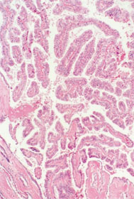
A: |
A papillary structure with atypical cells and the proliferation of tumor cells is evident (papillary carcinoma). |
|
|
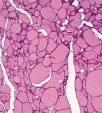
B: |
Normal thyroid tissue (colloid follicles show a regular arrangement). |
|
|
C: |
A nodular tumor is evident in the cervical region. |
|
|
|
|
|
|
|
|
|
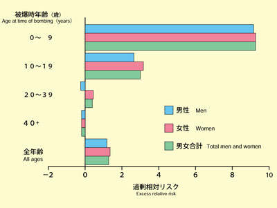
D: Excess relative risk of thyroid cancer(1958-1987) |
|
|
|
 |
|
|
|
|
|
|
• |
The increase in excess relative risk at 1 Sv for all ages was 1.15. |
|
|
• |
The excess relative risk was higher in people who were young at the time of bombing than in those who were old at the time of bombing.
[Thompson DE, et al., Rediat Res 137:S17-S67, 1994] |
|
|
|
|
|
|
|
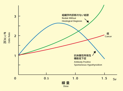
E: Thyroid disease in the Nagasaki atomic bomb survivors |
|
|
|
 |
|
|
|
|
|
|
• |
Solid nodules of the thyroid (thyroid cancer, adenoma, adenomatous goiter, nodules without histological diagnosis) were significantly more frequent among the atomic bomb survivors than among non-exposed persons, and it is known that the number of cases was higher the higher the dose and the lower the age at the time of bombing. |
|
|
• |
Antibody-positive spontaneous hypothyroidism was significantly more frequent among the atomic bomb survivors than among non-exposed persons. Moreover, the frequency reached a peak at a dose of 0.7Sv, which was lower than that in cancer cases.
[Cited and modified from Nagataki S, et al., JAMA 272:364-370, 1994] |
|
|
|
|
|